Kidney stones are a common urological condition that can cause severe pain and discomfort if not treated on time. These stones form due to the buildup of minerals and salts in the kidneys and can affect people of all age groups.
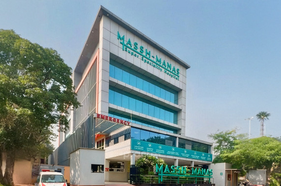
If you are looking for kidney stone treatment in Noida, it is important to choose a hospital that offers accurate diagnosis and advanced treatment options. At MASSH-MANAS Super Speciality Hospital, Noida, we provide comprehensive kidney stone care using modern techniques such as laser lithotripsy, RIRS, URS, and PCNL.
Our goal is to deliver effective, minimally invasive treatment that ensures faster recovery and long-term relief.
If you are experiencing symptoms such as severe back pain, blood in urine, or difficulty urinating, consult the expert urologists at MASSH-MANAS Super Speciality Hospital, Noida.
We offer advanced treatment options including laser lithotripsy, RIRS, URS, and PCNL for safe and effective stone removal.
📞 Call Now: +91-7290975999
📅 Book Appointment Online

Kidney stones are hard deposits formed from minerals like calcium, oxalate, and uric acid. These stones develop when urine becomes concentrated, allowing crystals to stick together and grow in size.
They can remain in the kidneys or travel through the urinary tract, causing varying levels of discomfort. The urinary system—including kidneys, ureters, bladder, and urethra—may be affected during this process.
There are several types of kidney stones, classified based on their composition:
These are the most prevalent type, responsible for nearly 80% of all kidney stones. They usually consist of calcium oxalate linked to high dietary oxalate or increased oxalate production in the body or calcium phosphate, which tends to develop in people with metabolic imbalances or persistently alkaline urine.
Forming in highly acidic urine, these stones are often associated with diets rich in animal protein, insufficient hydration, or medical issues like gout. They represent around 10% of all kidney stone cases.
Less frequently seen, struvite stones are a result of urinary tract infections caused by bacteria that generate ammonia. These stones can enlarge rapidly and sometimes take on a branched, "staghorn" shape.
These rare stones arise in individuals with a genetic disorder known as cystinuria, where excessive amounts of the amino acid cystine are released into the urine by the kidneys.
Diagnosis begins with a detailed clinical evaluation. Doctors may recommend:
These tests help determine the size, location, and type of kidney stone, enabling doctors to plan the most effective treatment.
| Severity | Stone Size | Treatment Approach |
|---|---|---|
| Mild | <5 mm | Medication & hydration |
| Moderate | 5–10 mm | RIRS / Laser treatment |
| Severe | >10 mm | PCNL / Surgery |
At MASSH-MANAS Super Speciality Hospital, we offer complete and advanced kidney stone treatment in Noida tailored to patient needs.
Patients from Noida, Greater Noida, Sector 62, Sector 18, Indirapuram, and Ghaziabad regularly visit us for expert care.
Small stones may pass naturally with:
👉 Learn more about related urology care:
Laser lithotripsy is one of the most advanced and commonly used treatments.
A flexible scope is used to access and treat kidney stones internally.
Recommended for large or complex stones.
Used for stones located in the ureter.
👉 Get advanced kidney stone treatment with minimally invasive techniques.
For accurate cost, consultation is recommended.
Patients from Noida, Greater Noida, Sector 62, Sector 18, Indirapuram, and Ghaziabad trust our expertise for kidney stone care.
Recovery depends on the treatment method used.
| Foods to Avoid | Reason |
|---|---|
| High salt foods | Increase calcium excretion |
| Spinach | High oxalate content |
| Red meat | Raises uric acid |
| Recommended Foods | Benefit |
|---|---|
| Citrus fruits | Prevent stone formation |
| Water | Flushes toxins |
| Vegetables | Maintain balance |
Consult a doctor if you experience:
MASSH-MANAS Urology Team
Senior Consultants – Urology
MASSH-MANAS Super Speciality Hospital, Noida
Our team of experienced urologists has over 20 years of expertise in performing advanced kidney stone procedures, including laser lithotripsy, RIRS, URS, and PCNL, with a focus on safety, precision, and optimal patient outcomes.
Disclaimer: This content is for educational purposes only and does not replace professional medical consultation.
Embark on a journey of exceptional healthcare guided by industry's true luminaries who consistently exceed
expectations and set new benchmarks for excellence in everything from cutting-edge innovations to personalized care.

MASSH MANAS Hospital distinguishes itself in kidney stone treatment through its advanced technology, highly skilled doctors, 3D laparoscopy, and patient-centric environment:
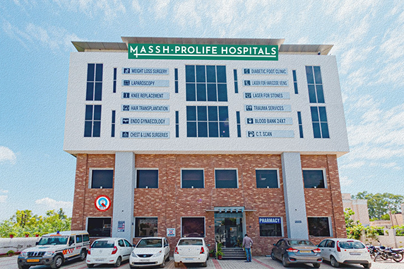
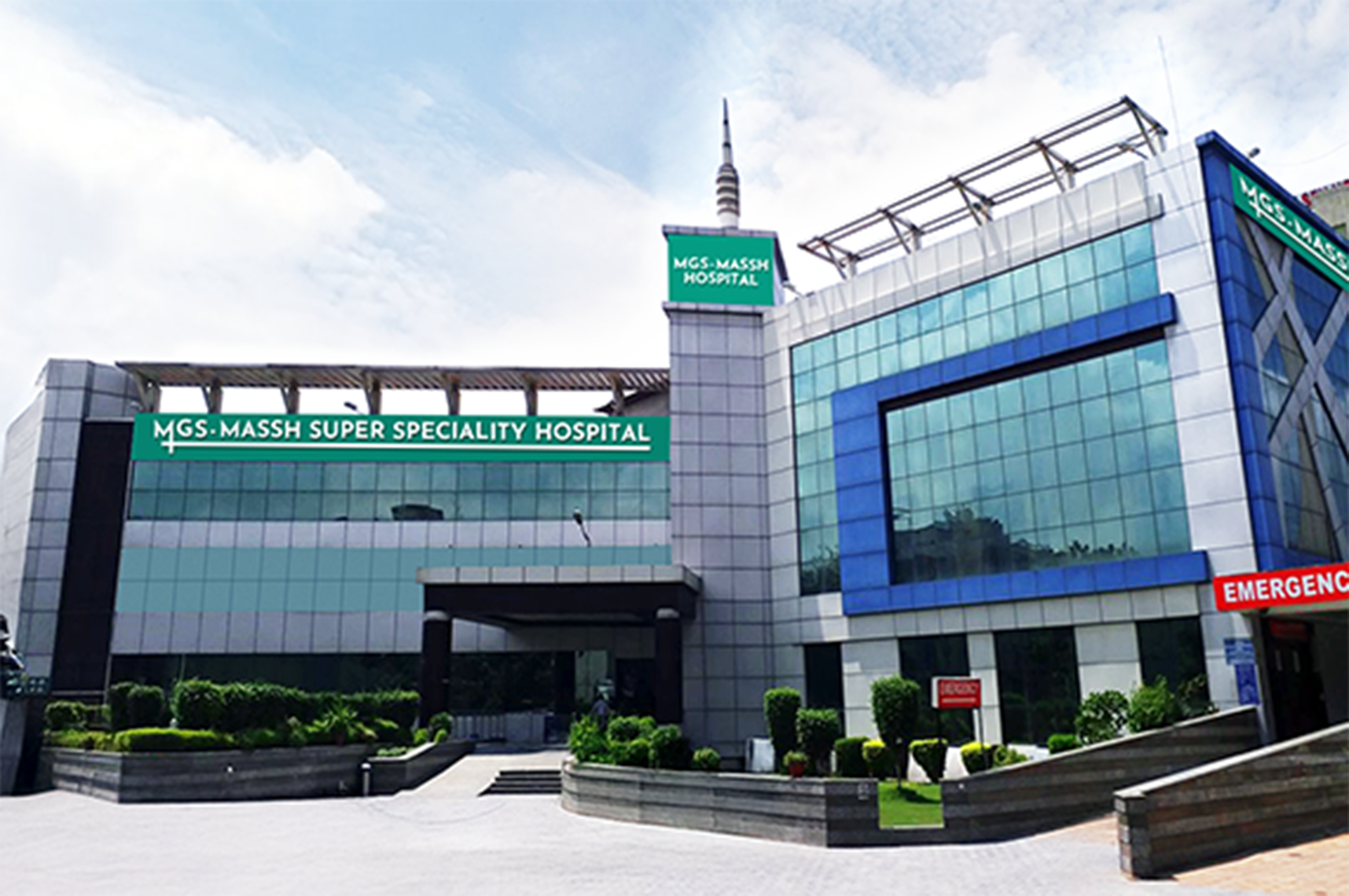
MASSH Group of Hospitals is a trusted name in delivering advanced healthcare solutions with a network of premier super speciality hospitals, committed to providing ethical, compassionate, and innovative care. Our expanding network ensures that cutting-edge medical services are always within your reach.
Kidney stones form due to mineral buildup caused by dehydration, diet, or metabolic conditions.
No, small stones may pass naturally. Surgery is needed for larger stones.
Modern treatments are minimally invasive and designed to reduce discomfort.
Yes, it is a minimally invasive and widely used procedure.
Most patients recover within a few days.
Depends on your condition:
Post-treatment care varies by method (natural passage, lithotripsy, surgery), but includes:
Yes, recurrence is possible without lifestyle changes.
Yes, it is widely used and safe.