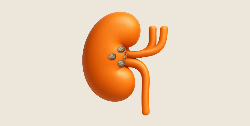
Renal stones, commonly known as kidney stones or nephrolithiasis, are dense mineral deposits that develop inside the kidneys. They result from high concentrations of substances like calcium, oxalate, uric acid, or phosphate in the urine, which form crystals that grow into stones. These may range in size and can stay in the kidney or move through the urinary tract, leading to sharp pain and discomfort depending on their location.
Types of Kidney Stones
There are several types of kidney stones, classified based on their composition:
Calcium Stones: Accounting for roughly 80% of kidney stones, these are the most frequently encountered type. They typically form as calcium oxalate stones linked to high oxalate levels in the diet or body or as calcium phosphate stones, which are often connected to metabolic disorders or elevated urine pH.
Uric Acid Stones: These stones develop when urine becomes overly acidic. Common causes include diets high in red meat, inadequate fluid intake, and conditions such as gout. They make up about 10% of all kidney stones.
Struvite Stones: These less common stones result from urinary tract infections caused by specific bacteria that produce ammonia. Struvite stones can expand rapidly and may take the shape of “staghorn” formations filling parts of the kidney.
Cystine Stones: A rare form of stone, cystine stones occur in individuals with cystinuria a hereditary disorder that causes the kidneys to excrete too much of the amino acid cystine into the urine.
Kidney Stones : Signs and Symptoms
Kidney stones may not cause symptoms until they move within the kidney or pass into the ureter (the tube connecting the kidney to the bladder). When symptoms occur, they can include:
Severe Pain: Sharp or cramping pain in the lower back, side, abdomen, or groin, often described as one of the most intense pains a person can experience. The pain may come in waves and shift as the stone moves.
Blood in Urine (Hematuria): Urine may appear pink, red, or brown due to irritation or damage to the urinary tract.
Nausea and Vomiting: Caused by the intense pain or a blockage affecting kidney function.
Frequent Urination or Urgency: A sensation of needing to urinate more often, sometimes with only small amounts passed.
Painful Urination: Burning or discomfort when urinating, especially if the stone is near the bladder.
Fever and Chills: If an infection develops alongside the stone, indicating a potentially serious complication.
Small stones may pass unnoticed, but larger ones can cause blockages, leading to swelling of the kidney (hydronephrosis) or infection.
Kidney Stones Causes
Dehydration: Not drinking enough water is a key factor in kidney stone development. When dehydrated, the body produces less urine, which becomes concentrated. This limits the urine’s ability to keep minerals such as calcium, oxalate, and uric acid dissolved, allowing them to clump and crystallize. Those in hot regions or who sweat a lot without fluid replacement are at higher risk. Chronic dehydration worsens this by creating a constant setting for stone-forming materials to accumulate.
Metabolic Imbalances: Kidney stones may arise from irregularities in mineral metabolism. Hypercalciuria, for example, involves excessive urinary calcium due to overactive parathyroid glands controlling calcium levels. Uric acid stones can form when uric acid is elevated, often linked to gout or purine breakdown, particularly in acidic urine conditions.
Urinary Stasis: Any condition slowing or blocking urine flow can lead to stones. Problems like an enlarged prostate, kidney abnormalities (e.g., horseshoe kidney), or obstructions in the urinary tract cause minerals to stagnate and crystallize instead of being flushed away.
Infections: Certain urinary infections, particularly from bacteria like Proteus or Klebsiella, can trigger struvite stones. These infections make urine more alkaline, encouraging magnesium, ammonium, and phosphate to form stones, known as "infection stones," which can enlarge rapidly.
Kidney Stones Risk Factors
Sodium: Excessive salt intake (think processed foods, salty snacks, or heavy seasoning) increases calcium excretion in the urine. More calcium in the urine means a higher chance it’ll bind with oxalate or phosphate to form stones.
Animal Protein: Diets heavy in meat, poultry, or fish (rich in purines) boost uric acid production and acidify the urine, creating a double whammy for stone formation. Protein also increases calcium and oxalate levels in urine while reducing citrate—a natural stone inhibitor.
Oxalate-Rich Foods: Foods like spinach, rhubarb, beets, nuts, chocolate, and even tea contain high levels of oxalate. When oxalate combines with calcium in the urine, it forms calcium oxalate stones, the most common type. People who over consume these foods without balancing them with adequate hydration or calcium from other sources (like dairy) are at greater risk.
Low Fluid Intake: Tied to dehydration, consistently drinking too little water (less than 2-3 liters daily for most people) is a major risk factor. Urine output below 1 liter per day significantly raises stone risk, as there’s simply not enough liquid to flush out stone-forming substances.
Family or Personal History: Genetics play a role. If your parents or siblings have had kidney stones, your odds go up—possibly due to inherited metabolic tendencies like hypercalciuria or low citrate production. A past stone also makes recurrence more likely, especially without lifestyle changes.
Obesity: Excess body weight alters urine chemistry, increasing calcium, oxalate, and uric acid levels while decreasing pH (making urine more acidic). Studies show obese individuals have a higher incidence of stones, partly due to insulin resistance affecting mineral metabolism.
Medical Conditions: Certain diseases amplify risk. Gout increases uric acid stones. Inflammatory bowel diseases (like Crohn’s) or gastric bypass surgery disrupt oxalate absorption, flooding the urine with it. Type 2 diabetes, hypertension, and chronic kidney disease also tweak urine composition in stone-friendly ways.
Medications and Supplements: Overuse of vitamin C supplements (which convert to oxalate), laxatives, or calcium-based antacids can tip the scales toward stone formation. Some diuretics or protease inhibitors (used in HIV treatment) also contribute.
Climate and Lifestyle: Living in hot, dry regions or working in jobs with heavy sweating like construction ups the risk if fluid intake doesn’t match output. Sedentary lifestyles may also indirectly contribute by slowing metabolism and urine flow.
Complications of Kidney Stones
Kidney stones can lead to serious issues if untreated:
Urinary Blockage: When a stone obstructs the ureter, it can halt urine passage, leading to kidney swelling (hydronephrosis) and possibly long-term kidney injury.
Infections: Stagnant urine behind a blockage can become a breeding ground for bacteria, increasing the risk of urinary tract infections or even life-threatening sepsis.
Intense Discomfort: Severe, unrelenting pain caused by kidney stones can interfere with daily functioning and may require urgent medical intervention.
Kidney Function Decline: Repeated episodes or ongoing obstruction from stones can gradually damage the kidneys and reduce their efficiency.
Bleeding in Urine: As stones move through the urinary tract, they may irritate or injure delicate tissues, resulting in visible or microscopic blood in the urine (hematuria), and in rare cases, notable blood loss.
Kidney Stone Diagnosis
Medical History Review:
The doctor asks about symptoms (e.g., pain, urination issues), past stone history, diet, and family history of kidney stones.
Physical Examination:
Checks for pain in the back, sides, or abdomen; assesses general health to rule out other conditions.
Urinalysis:
Urine sample tested for blood, crystals, infection, or abnormal levels of stone-forming substances (e.g., calcium, uric acid).
Blood Tests:
Measures levels of calcium, uric acid, or other markers to identify underlying metabolic issues or kidney function.
Imaging Tests:
Ultrasound:
CT Scan:
X-ray (KUB):
Stone Analysis (if passed):
Examines a passed stone’s composition (e.g., calcium oxalate, uric acid) to guide treatment and prevention.
About Kidney Stone Size
Kidney stones are sized in millimeters (mm), and their size plays a major role in whether they pass on their own or need treatment. Tiny stones (1–4 mm, like grains of sand) usually pass unnoticed with good hydration. Stones measuring 4–6 mm (about the size of a sesame seed) have an 80% chance of passing, though they can cause pain. Stones around 5–6 mm pass in about 50–60% of cases and may require medications. Larger ones (6–10 mm) pass in less than 20% of cases, often needing RIR. Stones 10 mm and above rarely pass and typically need PCNL. Factors like size, type, and location matter.
Kidney Stone Treatment Options
Treatment depends on the stone’s size, type, location, and severity of symptoms. Options include:
Conservative Management:
Hydration: Drinking plenty of water (8-12 cups daily) to help small stones (less than 4-6 mm) pass naturally.
Pain Relief: Over-the-counter medications like ibuprofen or prescription drugs to manage discomfort.
Medical Expulsive Therapy (MET): Medications like tamsulosin to relax the ureter and facilitate stone passage.
Minimally Invasive Procedures:
RIRs surgery: This is done using a thin, flexible instrument called a ureteroscope that is inserted through the urethra and bladder to reach the kidney or the ureter.
Ureteroscopy: A thin scope is inserted through the urethra to locate and break up stones with a laser. Fragments are removed or passed naturally.
Percutaneous Nephrolithotomy (PCNL): For larger stones (>2 cm), a small incision is made in the back, and a scope is used to remove or break up the stone. Requires a short hospital stay.
MiniPerc (Minimally Invasive Percutaneous Nephrolithotomy): MiniPerc, or Minimally Invasive Percutaneous Nephrolithotomy, is a procedure that removes kidney stones via a small cut in the skin, through which precise instruments access the kidney. Using smaller equipment and a tinier incision than conventional PCNL, it causes less physical strain, promotes faster healing, and reduces complications.
Surgical Intervention:
Open Surgery: Rarely used today, reserved for very large or complex stones when other methods fail. It involves a larger incision and longer recovery.
Prevention: After treatment, dietary changes (e.g., reducing sodium, animal protein, or oxalate-rich foods) and medications (e.g., thiazide diuretics for calcium stones or allopurinol for uric acid stones) may be recommended to prevent recurrence.
Embark on a journey of exceptional healthcare guided by industry's true luminaries who consistently exceed expectations and set new benchmarks for excellence in everything from cutting-edge innovations to personalized care.
Why You Should Not Ignore Treatment for Kidney Stones
Can lead to severe pain – Untreated stones often cause intense, recurring pain.
Risk of kidney damage – Prolonged blockage can impair kidney function.
Urinary tract infections (UTIs) – Stones can increase the risk of infections.
Stone growth and recurrence – Delayed treatment may lead to larger or more stones.
Emergency complications – Ignoring stones may result in emergency situations like hydronephrosis.
Care at MASSH - PROLIFE
Mash Hospital distinguishes itself in kidney stone treatment through its advanced technology, highly skilled doctors, 2D laparoscopy, and patient-centric environment:
Advanced Technology: At MASSH – PROLIFE Hospital, we rely on modern diagnostic systems like detailed CT imaging and ultrasound, paired with latest-generation equipment for accurate diagnosis and treatment. This approach allows for precise stone targeting and removal, minimizing complications and improving success rates while enhancing overall patient safety and care.
Highly Skilled Doctors: The hospital boasts a team of experienced urologists and nephrologists who specialize in kidney stone management. Their expertise allows for tailored treatment plans, from non-invasive options to complex surgeries, ensuring optimal outcomes.
2D Laparoscopy: Unlike traditional 2D methods, 2D laparoscopy provides surgeons with enhanced depth perception and precision during minimally invasive procedures like PCNL or ureteroscopy. This reduces operative time, improves accuracy, and speeds up recovery.
Home-Like Environment: MASSH PROLIFE Hospital prioritizes patient comfort with a welcoming, supportive atmosphere. This includes personalized care, comfortable recovery spaces, and emotional support, making the treatment experience less stressful and more reassuring.
Cashless Treatment Facility: At MASSH – PROLIFE Hospital in Ludhiana, we offer convenient cashless treatment for patients insured under associated TPAs and insurance firms. To use this facility, patients must provide essential paperwork including a photo ID, valid mediclaim card or policy, a prescription indicating admission, and a duly filled TPA form. All documents must be submitted to the Insurance Desk within 24 hours of hospital entry. The hospital then contacts the TPA to start the pre-authorization. Final approval generally follows 3–4 hours after discharge billing and summary submission. Covered expenses are paid by the insurer up to the approved amount. Patients should confirm if their insurer is on our panel and verify cashless eligibility in advance. Non-approved costs will need to be paid by the patient. Our Insurance Desk coordinates efficiently to ease the patient’s financial concerns.
HOSPITALS
MASSH Group of Hospitals is a trusted name in delivering advanced healthcare solutions with a network of premier super speciality hospitals, committed to providing ethical, compassionate, and innovative care. Our expanding network ensures that cutting-edge medical services are always within your reach.
Ureteroscopy: A scope is used to remove or fragment stones in the ureter or kidney.
Percutaneous Nephrolithotomy (PCNL): Surgery via a small back incision for large stones (>10 mm).
Open Surgery: Rarely used, for complex cases.
Yes, many do, especially if <5 mm (50-80% chance). Stones 6-9 mm pass less often (<20%), and those ≥10 mm usually require intervention. Hydration and meds can aid passage.
1-4 mm: Days to 1-2 weeks.
5-6 mm: 2-4 weeks, if it passes.
Larger Stones: Often need treatment; time depends on method. Location, shape, and ureter size impact duration.
General Goal: 2.5-3 liters (85-100 oz) daily, aiming for ≥2 liters of urine output.
Stone Formers: 3-4 liters, adjusted for climate, activity, or doctor’s advice. Increase for sweating (e.g., heat, exercise).
Depends on symptoms and stone status:
Asymptomatic: Small, stable kidney stones allow light to moderate exercise (e.g., walking, yoga), which may aid urine flow.
Painful or Passing: Avoid vigorous activity (running, lifting) during pain or passage to prevent worsening discomfort or stone movement. Rest or light movement is better.
Post-Treatment: After lithotripsy or surgery, wait 1-2 weeks (or per doctor’s advice) before intense activity to avoid strain or bleeding.
Hydration Key: Drink water before, during, and after exercise to avoid dehydration, a stone trigger. Consult your doctor if you have pain, fever, or a large stone.
Care varies by treatment (natural passage, lithotripsy, surgery), but generally:
Hydrate Aggressively: Drink 2.5-3 liters daily to flush fragments and prevent new stones. Strain urine if advised to collect pieces for analysis.
Follow Medication Instructions: Take pain relievers, antibiotics (for infections), or preventive drugs (e.g., potassium citrate) as prescribed.
Rest as Needed: Avoid heavy lifting or strenuous activity for 1-2 weeks post-surgery or lithotripsy (per doctor’s guidance) to support kidney/ureter healing.
Watch for Complications: Contact your doctor if you have fever, severe pain, heavy bleeding, or urination issues—these may indicate infection or blockage.
Diet Adjustments: Begin prevention (low sodium, moderate protein) immediately to reduce recurrence risk.
Follow-Up: Get imaging or urine tests to ensure stones are cleared and kidneys are functioning. Discuss long-term prevention based on stone type. Healing takes days (natural passage) to weeks (procedures); staying proactive lowers risks.
AFFORDABLE & ACCESSIBLE
Centre of Excellence for
CANCER CARE
Get a call back for Free Consultation
Organising
Free Mega Breast Cancer
Screening Camp
From 1st to 31st October
Get a callback for Free Screening & Consultation
×
Download PDF!
×
Get An Instant Callback!
Register Now and Get Free Consultation by Senior Consultant.
×
Get a Free Consultation
Register now and get free consultation by a senior consultant.